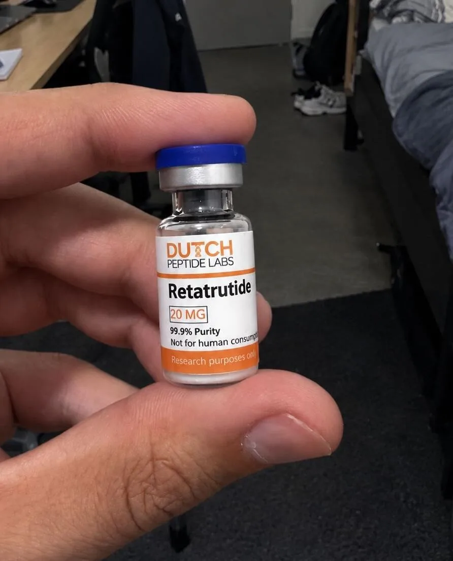





An intact mass determination by LC-MS confirms that a research peptide has the correct molecular weight, but says little about the exact order of the amino acids within it. Two peptides with identical mass can have entirely different sequences through a swap of residues. Peptide mapping is the technique that does make this distinction: it cuts a peptide into smaller fragments that are easy to analyse, and in doing so establishes the full sequence. For the quality control of research peptides it is a valuable complement to intact mass and purity analyses.
Why intact mass is not always sufficient
A mass spectrometer measures the molecular weight of an intact peptide with high accuracy, and in combination with the RP-HPLC retention time this gives a strong indication of identity. Even so, there are scenarios in which this offers insufficient certainty: isobaric amino acid substitutions (such as leucine and isoleucine, which have exactly the same mass), swaps between two residues that together give the same mass as another pair, or a deletion that happens to be compensated by another deviation. In these cases the intact mass shows no sequence error, while the actual amino acid order differs from the intended sequence. Peptide mapping was developed to detect precisely this kind of hidden deviation by examining the peptide at sequence level rather than as a single whole.
Enzymatic and chemical digestion: cutting up the peptide
The first step in peptide mapping is fragmenting the research peptide at predictable sites. Enzymatic digestion with proteases such as trypsin (cleaves after lysine and arginine) or Glu-C (cleaves after glutamic acid) is the most widely used approach, because these enzymes cut in a highly specific and reproducible way. For peptides that contain no suitable trypsin or Glu-C sites, or for targeted structure elucidation, chemical digestion is sometimes used instead, such as cleavage with cyanogen bromide (CNBr) at methionine residues. The choice of digestion method depends strongly on the amino acid composition of the specific research peptide: a sequence without lysine or arginine calls for a different enzyme or a chemical route than a lysine-rich peptide.
Fragment analysis with LC-MS/MS
After digestion, the fragment mixture is separated by reversed-phase HPLC coupled to tandem mass spectrometry (LC-MS/MS). Each fragment is first weighed (MS1) and then fragmented further within the instrument (MS2), with the resulting collision fragments revealing the amino acid order within that piece of peptide. By comparing the measured fragment masses with the theoretically expected fragments of the intended sequence, a “sequence coverage” is obtained: the percentage of the sequence that has actually been confirmed experimentally. Full or near-full coverage gives considerably more certainty about sequence integrity than a single intact mass measurement alone can offer.
What peptide mapping adds to research QC
In the context of quality control for research peptides, peptide mapping is mainly used as orthogonal confirmation alongside the usual combination of RP-HPLC purity and intact mass spectrometry, particularly for longer sequences, peptides with several modified residues, or when earlier analyses gave an ambiguous result. Published methods literature describes peptide mapping via chemical digestion and MALDI-MS as an established approach to sequence confirmation in protein and peptide characterisation, and the same principles apply to synthetic research peptides. For routine batch release the method is more labour-intensive than a standard CoA check, which is why it is more often reserved for targeted identity confirmation than used for every batch produced.
Practical considerations in interpretation
Incomplete sequence coverage does not automatically mean a sequence error: some fragments are too small or too hydrophilic to be retained well on the HPLC column, and may therefore be lost before they reach the mass spectrometer. Researchers who apply peptide mapping or have it carried out take this into account by tailoring the digestion strategy to the specific sequence and by combining several enzymes when a single enzyme gives insufficient coverage. This produces a more complete and more reliable picture of the actual amino acid order of the research material.
The products and information on this website are intended solely for laboratory and research purposes. They are not intended for human or animal consumption, nor for diagnostic or therapeutic use.